
Regulation of Snf1 kinase: activation requires phosphorylation of threonine 210 by an upstream kinase as well as a distinct step mediated by the Snf4 subunit |
Department of Molecular Genetics and Biochemistry
University of Pittsburgh School of Medicine
Pittsburgh, PA 15261
| The yeast Snf1 kinase and its metazoan orthologues, the AMP-activated protein kinases, are activated in response to nutrient limitation. Activation requires the phosphorylation of a conserved threonine residue in the activation loop of the catalytic subunit. A phosphopeptide antibody was generated that specifically recognizes Snf1 protein that is phosphorylated in its activation loop on threonine 210. Using this reagent, we show that phosphorylation of threonine 210 correlates with Snf1 activity since it is detected in cells subjected to glucose limitation but not in cells grown in abundant glucose. A Snf1 mutant completely lacking kinase activity was phosphorylated normally on threonine 210 in glucose starved cells, eliminating the possibility that the threonine 210 modification is due to an auto-phosphorylation event. Cells lacking the Reg1 protein, a regulatory subunit for the Glc7 phosphatase, showed constitutive phosphorylation of Snf1 threonine 210. Exposure of cells to high concentrations of sodium chloride also induced phosphorylation of Snf1. Interestingly, Mig1, a downstream target of Snf1 kinase, is phosphorylated in glucose stressed but not sodium stressed cells. Finally, cells lacking the gamma subunit of the Snf1 kinase complex encoded by the SNF4 gene exhibited normal regulation of threonine 210 phosphorylation in response to glucose limitation but are unable to phosphorylate Mig1 efficiently. Our data indicate that activation of the Snf1 kinase complex involves two steps, one that requires a distinct upstream kinase and one that is mediated by the gamma subunit of the kinase itself. |
|
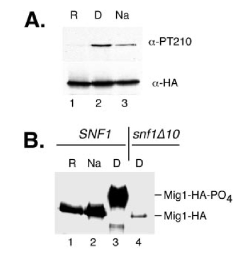
Phosphorylation of Thr210 in response to sodium ion stress. A. Western blot of Snf1 protein collected by immunoprecipitation with anti-HA antibodies from 300 ug of protein. The upper panel shows reactivity with anti-threonine 210 antibodies (-PT210), and the lower panel shows reactivity with anti-HA antibodies (-HA). Extracts were prepared from cells grown under repressing conditions (R; lane 1), derepressing conditions (D; lane 2) or in repressing conditions supplemented with 0.8 M NaCl for 1 h (Na; lane 3). B. Western blot of Mig1 protein. Extracts were prepared from cells expressing HA-tagged Mig1 protein that had been grown under repressing conditions (R; lane 1), derepressing conditions (D; lanes 3 and 4), or repressing conditions supplemented with 0.8 M NaCl for 1 h (Na; lane 2). Cells were wild type 10 (lane 4). Each lane contained 80 ug of protein. |