| front |1 |2 |3 |4 |5 |6 |7 |8 |9 |10 |11 |12 |13 |14 |15 |16 |17 |18 |19 |20 |21 |22 |23 |24 |25 |26 |27 |28 |29 |30 |31 |32 |33 |34 |35 |36 |37 |38 |39 |40 |41 |42 |43 |44 |45 |46 |47 |48 |49 |50 |51 |52 |53 |54 |55 |56 |review |
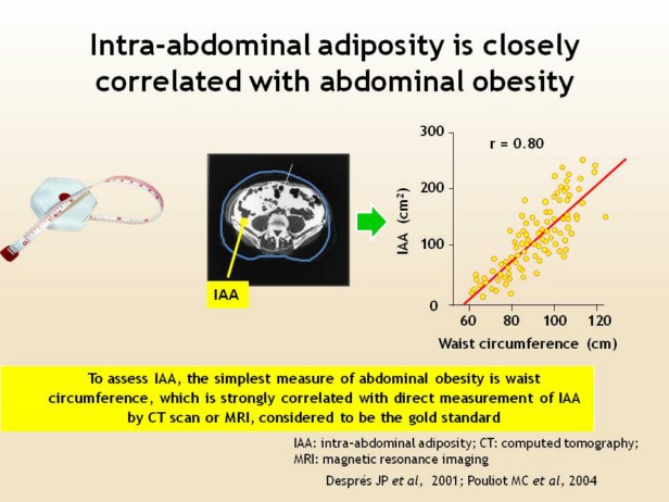 |
Here we see an example of a CT scan in a patient with intra-abdominal adiposity, showing the accumulation of fat around the visceral organs. CT scanning is considered to represent the ‘gold standard’ for measurement of body fat distribution, but is too complex and costly for routine use for this purpose. Abdominal obesity, as measured by waist circumference, correlates closely with intra-abdominal adiposity measured using CT scanning. The data shown on this slide are from a population of 81 men and 70 women. Abdominal obesity, with diagnostic criteria based on waist circumference, is a requirement for the diagnosis of the metabolic syndrome according to the new guidelines from the International Diabetes Federation. This simple, straightforward and well-known measure should be adopted as part of standard clinical practice for diagnosing intra-abdominal adiposity which, in turn, signifies increased cardiovascular risk.
|