| front |1 |2 |3 |4 |5 |6 |7 |8 |9 |10 |11 |12 |13 |14 |15 |16 |17 |18 |19 |20 |21 |22 |23 |24 |review |
 |
Depolarization results in a
failure to maintain ionic gradients and the normal hyperpolarized membrane potential. The
cells of the presynaptic membrane depolarize and release a flood of neurotransmitters, i.e.
glutamate. This depolarization is mediated by voltage-sensitive Na+
channels that carry electrical messages to the synapse. As a result of depolarization,
there is an influx of Ca2+ via the voltage-sensitive channels and a concomitant
secretion of glutamate ions. Although the principal targets of these neurotransmitters are
situated on the postsynaptic membrane, a number of presynaptic excitatory amino acid (EAA)
receptors and glutamate transporters receive the chemical message and respond in various
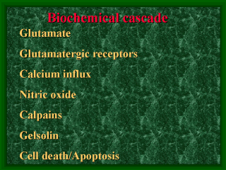
ways in order to regulate the actions of these messengers. (Dunlap et al. 1995) The accumulation of glutamate and the excessive activation of glutamate receptors mediate neuronal injury and cell death. As glutamate is highly neurotoxic in the extracellular space, it is continually being cleared in a normal environment, i.e. it is recycled by neuronal uptake through re-storage in the vesicles and re-release. Glial cells contain a similar, high-affinity, active transport that ensures the efficient removal of glutamate from the synapse. Ischaemia impairs the ability of cells to maintain such a glutamate uptake system. (Rhoades and Tanner 1995) Most of the excitotoxicity is related to the destructive actions of high intracellular calcium mediated by stimulation of the various subtypes of glutamatergic receptors. Glutamate receptors (Glur) are either ionotropic or metabotropic. The ionotropic receptors are ligand-gated ion channels which are further divided into three groups on the basis of which ligand preferentially activates the receptors: those that respond to N-methyl-D-aspartate (NMDA), -amino-3-hydroxy-5-methyl-4-isoxazoleproprionate (AMPA), and kainate (Small and Buchan 1996). The metabotropic receptors activate intracellular second messengers via G-proteins. (Pin and Duvoisin 1995) Massive release of glutamate and activation of glutamatergic receptors leads to Ca2+ influx, which starts numerous secondary processes that accelerate ischaemic neuronal damage. NO acts as a neuromodulator in the central nervous system and participates in many physiological processes such as brain development, pain perception, neuronal plasticity, memory function and behaviour. There are three isoforms of NO present in the brain: neuronal, endothelial and inducible. When overproduced, however,NO reverts from a neuromodulator to a neurotoxic mediator. Such an overproduction of NO may be a consequence of persistent stimulation of EAA receptors, but it can also be driven by cytokines. NO reacts with superoxide anions to generate the toxic substance peroxynitrite (ONOO–), which in turn oxidates proteins, lipids and DNA. This reaction can produce even more potent neurotoxins, such as hydroxyl radicals. In addition, ONOO– can disturb the glutamate transport system and possibly impair the mitochondrial respiratory chain. (Chabrier et al. 1999, Garthwaite et al. 1989). NO is also a potent dilator of the cerebral blood vessels. (Faraci and Brian 1994). Calpains are cytosolic proteinases whose proteolytic activity is directed mainly against the cytoskeleton and regulatory proteins. Calpains are activated by an increase in intracellular Ca2+, and their proteolysis produces irreversible degradation of a number of structural and regulatory proteins. (Bartus et al. 1998) High levels of intracellular Ca2+ activate gelsolin, a ubiquitous enzyme that severs actin microfilaments, thereby inhibiting any further influx of Ca2+ mediated by NMDA receptor-gated and voltage-gated Ca2+channels and acting as a neuroprotective factor (Endres et al. 1999). On the other hand, recent evidence suggests that gelsolin may promote apoptosis by reducing Ca2+ influx in instances of Ca2+ starvation. Gelsolin also seems to serve as a substrate for the critical apoptosis enzyme caspase. (Zipfel et al. 1999) There is an increasing amount of evidence that severe ischaemic injury may lead immediately to high glutamate levels, acute Ca2+ overload and cell death, whereas neurons with milder injury undergo apoptosis. (Zipfel et al. 1999). Caspases have been shown to mediate apoptotic neuronal cell death. (Gottron et al. 1997). Apoptosis is an active process requiring metabolic energy and protein and ribonucleic acid synthesis. The apoptotic cell is characterized by shrinkage, nuclear collapse and the cleaving of chromatin into nucleosomal fragments, while the organelles retain their integrity and there is no associated inflammation. (Arends and Wyllie 1991) |
| front |1 |2 |3 |4 |5 |6 |7 |8 |9 |10 |11 |12 |13 |14 |15 |16 |17 |18 |19 |20 |21 |22 |23 |24 |review |