| The Michael Group University of Pittsburgh |
|---|
|
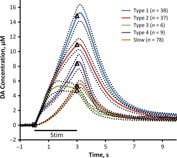
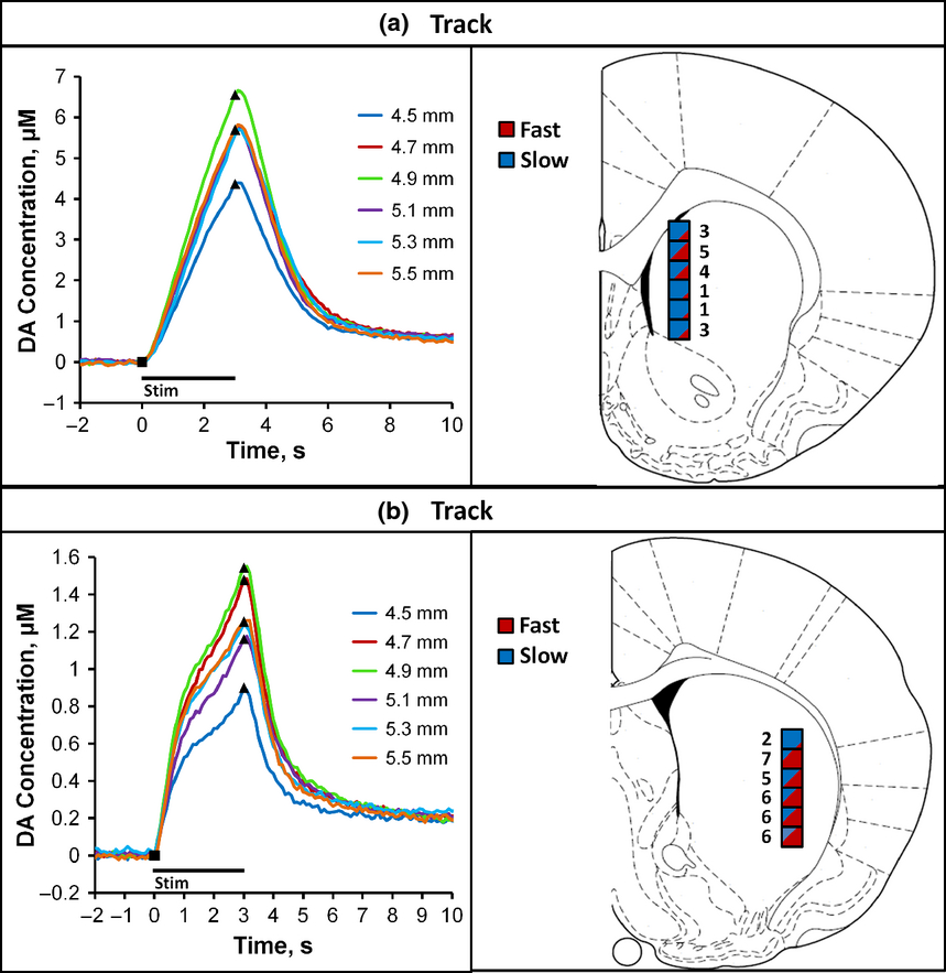
Kinetic diversity of dopamine transmission in the dorsal striatum Dopamine (DA), a highly significant neurotransmitter in the mammalian central nervous system, operates on multiple time scales to affect a diverse array of physiological functions. The significance of DA in human health is heightened by its role in a variety of pathologies. Voltammetric measurements of electrically evoked DA release have brought to light the existence of a patchwork of DA kinetic domains in the dorsal striatum (DS) of the rat. Thus, it becomes necessary to consider how these domains might be related to specific aspects of DA's functions. Responses evoked in the fast and slow domains are distinct in both amplitude and temporal profile. Herein, we report that responses evoked in fast domains can be further classified into four distinct types, types 1–4. The DS, therefore, exhibits a total of at least five distinct evoked responses (four fast types and one slow type). All five response types conform to kinetic models based entirely on first-order rate expressions, which indicates that the heterogeneity among the response types arises from kinetic diversity within the DS terminal field. We report also that functionally distinct subregions of the DS express DA kinetic diversity in a selective manner. Thus, this study documents five response types, provides a thorough kinetic explanation for each of them, and confirms their differential association with functionally distinct subregions of this key DA terminal field. The dorsal striatum is composed of five significantly different dopamine domains. Responses from each of these five domains exhibit significantly different ascending and descending kinetic profiles and return to a long lasting elevated dopamine state, termed the dopamine hang-up. All features of these responses are modeled with high correlation using first-order modeling as well as our recently published restricted diffusion model of evoked dopamine overflow. We also report that functionally distinct subregions of the dorsal striatum express selective dopamine kinetic diversity.
| |||||||

