| front |1 |2 |3 |4 |5 |6 |7 |8 |9 |10 |11 |12 |13 |14 |15 |16 |17 |18 |19 |20 |21 |22 |23 |24 |25 |review |
 |
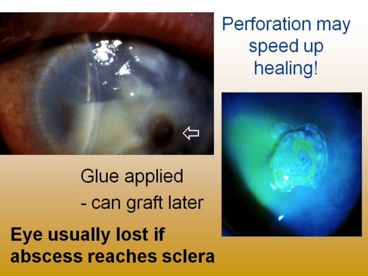
Left photo: brown iris protruding through perforation from infection; it plugs hole and brings vascular/immune system into play. Often will heal, and corneal transplant/iridoplasty can be done six months or more later, with reasonable outcomes. Right photo: cyanoacrylate glue with greenish fluorescein staining of adjacent corneal ulcer; soft contact lens placed over glue to reduce foreign body sensation. As lesion heals from inside, it will extrude glue in few months. Then consideration of corneal transplantation can be made. |